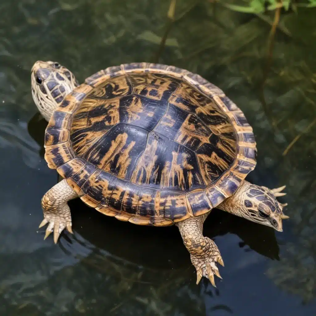
Performing an Electrocardiogram in Pond Sliders: A Comprehensive Guide
Reptilian Cardiac Monitoring
Reptiles, with their unique physiological adaptations, present both fascinating and complex challenges when it comes to cardiovascular health assessment. The pond slider turtle, or Trachemys scripta, is a popular exotic pet and conservation subject, making the development of reliable diagnostic tools a critical priority. Electrocardiography (ECG) stands out as a valuable non-invasive technique for evaluating cardiac function in these remarkable creatures.
Trachemys scripta Physiology
The pond slider turtle possesses a remarkable cardiovascular system tailored to its aquatic lifestyle and ectothermic nature. Unlike the four-chambered hearts of mammals, the chelonian heart is considered a tri-chambered or atypical four-chambered organ, with the sinus venosus functioning as a distinct chamber. This structural difference, combined with the unique electrical activation and conduction patterns, results in ECG waveforms that vary significantly from their mammalian counterparts.
The respiratory system of the pond slider also plays a crucial role in cardiovascular function. These turtles are capable of prolonged breath-holds, relying on both pulmonary and cutaneous gas exchange to meet their metabolic demands. Understanding the interplay between the respiratory and cardiac systems is essential when interpreting ECG findings in these animals.
Thermoregulation is another key aspect of pond slider physiology that can influence cardiac function. As ectotherms, these turtles rely on environmental temperatures to regulate their body temperature, which in turn impacts heart rate, conduction, and other ECG parameters.
Electrical Activity of the Heart
In contrast to the discrete sinoatrial node found in mammals, the electrical impulse in chelonians originates from cardiac muscle fibers within the sinus venosus. This electrical activation then spreads to the atria, followed by the atrioventricular junction and ventricular myocardium. The lack of specialized Purkinje fibers results in a unique pattern of ventricular depolarization, from base to apex, rather than the endocardial-to-epicardial progression seen in mammals.
Repolarization of the ventricular myocardium also differs, with the T wave often appearing similar to the QRS complex or R wave. The prolonged ventricular systole and rapid diastole characteristic of chelonians contribute to the distinct ECG waveform morphology and intervals observed in these species.
Electrode Placement
Obtaining high-quality ECG recordings in pond sliders requires strategic electrode placement to effectively capture the low-voltage electrical signals generated by the heart. Various non-invasive methods have been explored, each with its own advantages and limitations.
One of the most promising techniques involves applying adhesive patches to the prehumeral fossae and either the abdominal scutes of the plastron or the prefemoral fossae. This approach minimizes the obstruction caused by the thick plastron while positioning the electrodes in close proximity to the heart. Other methods, such as placing electrodes on the distal extremities or using alligator clips, have been described but may be more prone to motion artifact and reduced signal quality.
Signal Interpretation
Interpreting the ECG waveforms of pond sliders requires an understanding of the species-specific differences in cardiac electrical activity. The P wave, QRS complex, and T wave may exhibit variations in morphology, polarity, and duration compared to those observed in mammals. Additionally, the prolonged PR and QT intervals, as well as the absence of distinct Q and S waves, are characteristic features of the chelonian ECG.
Careful analysis of the ECG parameters, including heart rate, wave amplitudes, and intervals, can provide valuable insights into the cardiac health of pond sliders. Deviations from the established reference ranges may indicate underlying pathologies, such as arrhythmias, conduction disturbances, or myocardial dysfunction.
Turtle Handling Considerations
Performing ECG procedures in pond sliders requires a delicate balance of restraint, stress mitigation, and safety precautions to ensure the well-being of both the animal and the handler.
Restraint Techniques
Effective restraint is crucial to obtain high-quality ECG recordings without introducing motion artifact. The use of an inverted cup or similar device can provide a stable platform for the turtle, allowing it to remain conscious and minimally stressed throughout the procedure.
Stress Mitigation
Reducing the stress experienced by the turtle during the ECG process is essential to obtain reliable results and avoid compromising the animal’s health. Careful handling, a familiar environment, and the avoidance of excessive stimuli can help mitigate the stress response.
Safety Precautions
Working with reptiles, including pond sliders, requires specific safety considerations. Proper personal protective equipment, such as gloves, should be used to prevent the transmission of zoonotic diseases. Additionally, maintaining a controlled temperature environment and monitoring the turtle’s well-being throughout the procedure are crucial steps to ensure a safe and successful ECG session.
Methodology for ECG in Pond Sliders
The process of performing an ECG in pond sliders involves a series of carefully planned steps to ensure the collection of high-quality, reproducible data.
Preparation
Before the ECG procedure, the turtle should be acclimated to the environment and the handling protocol. Maintaining the appropriate temperature range, providing adequate lighting, and offering a familiar setting can help minimize the turtle’s stress response.
Data Collection
The ECG recording should be performed with the turtle in a stable, dorsal-ventral position, using adhesive patches or alligator clips strategically placed on the prehumeral fossae and the abdominal scutes of the plastron or the prefemoral fossae. A continuous 6-lead ECG tracing should be recorded for a minimum of 2 minutes, with the paper speed set at 50 mm/s and the sensitivity at 20 mm/mV.
Troubleshooting
Occasionally, challenges may arise during the ECG procedure, such as motion artifact or poor waveform recognition. In such cases, adjusting the lead placement, minimizing external stimuli, and ensuring the turtle’s comfort can help improve the quality of the recording.
By understanding the unique physiology of pond sliders, mastering the principles of ECG interpretation, and employing appropriate handling techniques, veterinary professionals and researchers can leverage this valuable diagnostic tool to enhance the care and management of these remarkable reptiles. The development and standardization of ECG methods for pond sliders represent an important step in advancing the field of reptile cardiology and improving the overall well-being of captive and wild populations.
For more information on avian and exotic animal care, I encourage you to visit the Mika Birds Farm website, where you can find a wealth of resources and expert guidance. Together, we can continue to expand our understanding and provide the best possible care for these unique creatures.


